Pancreatic Abscess in a 13-year-old, MN, Maine Coon Cat

Case Study

Pancreatic Abscess in a 13-year-old, MN, Maine Coon Cat

Anorexic, vomiting one day prior, lethargic.

PE: Firm mass palpated mid abdomen cranial to bladder. Seems uncomfortable on palpation. Given Cerenia, Convenia, Buprenorphine.

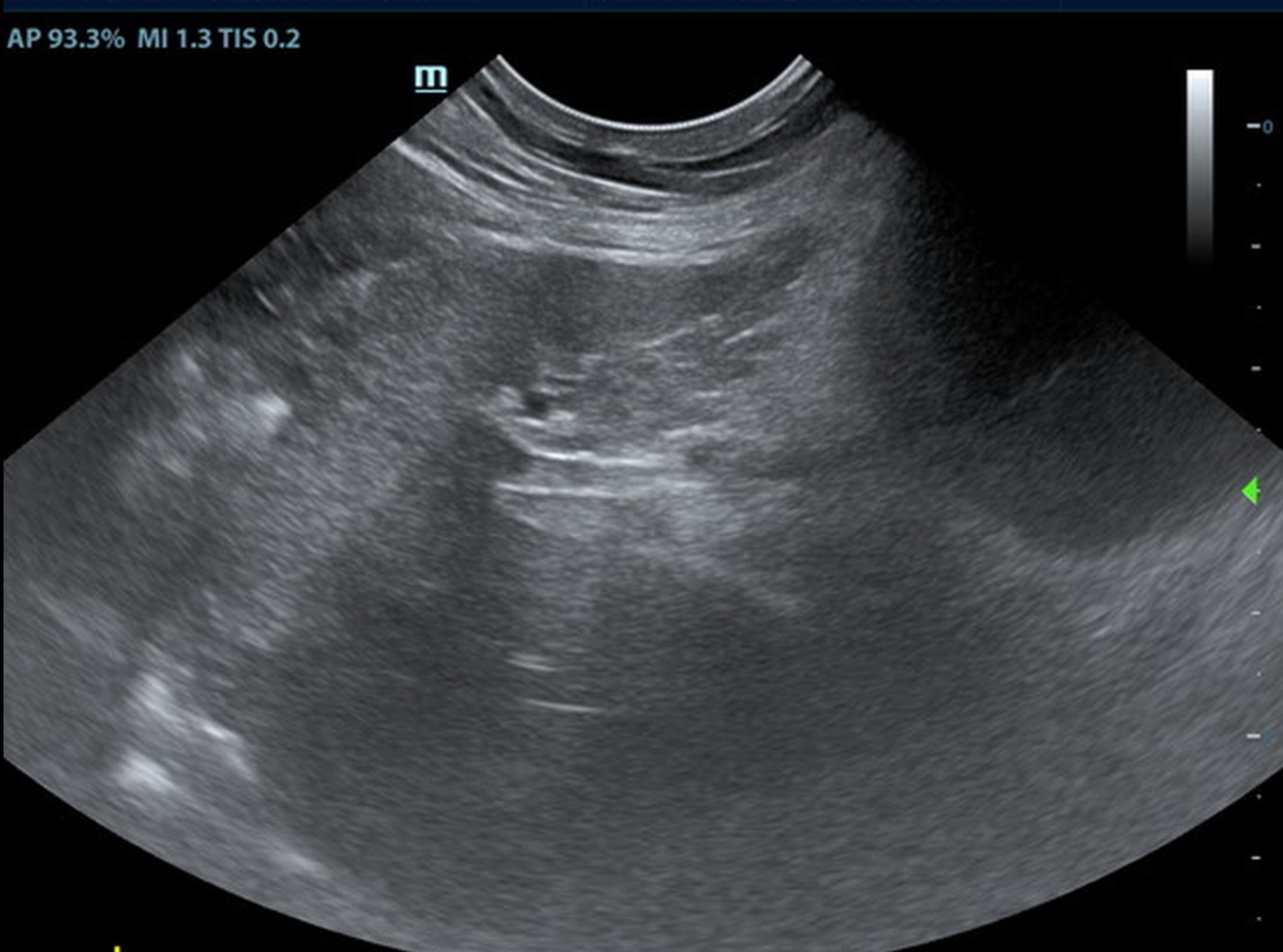
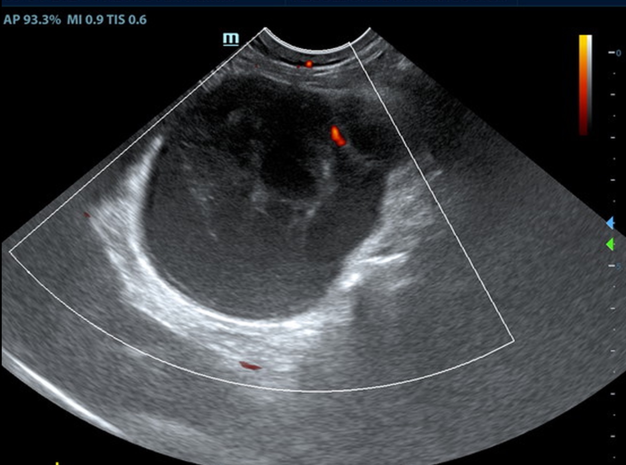
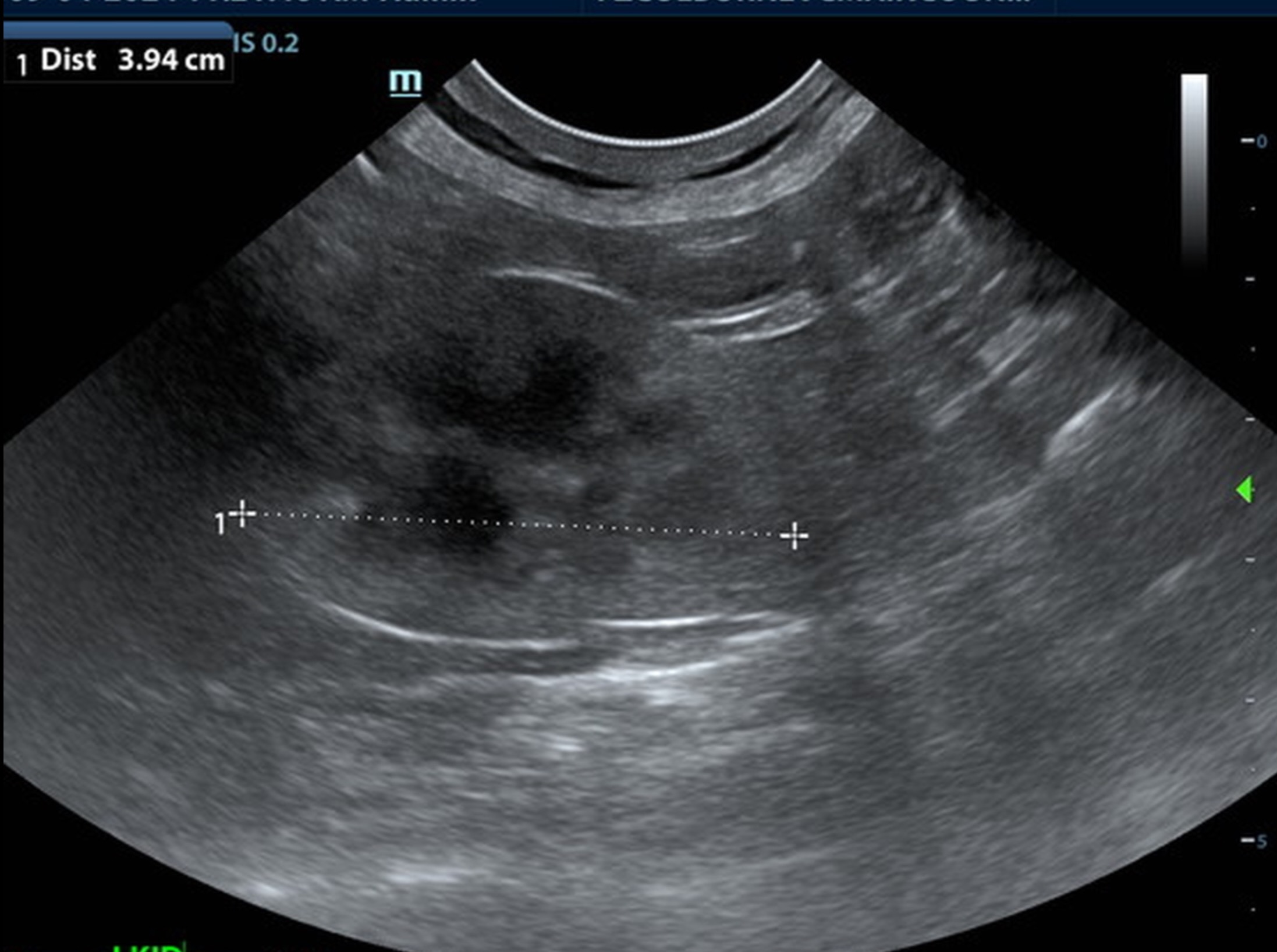
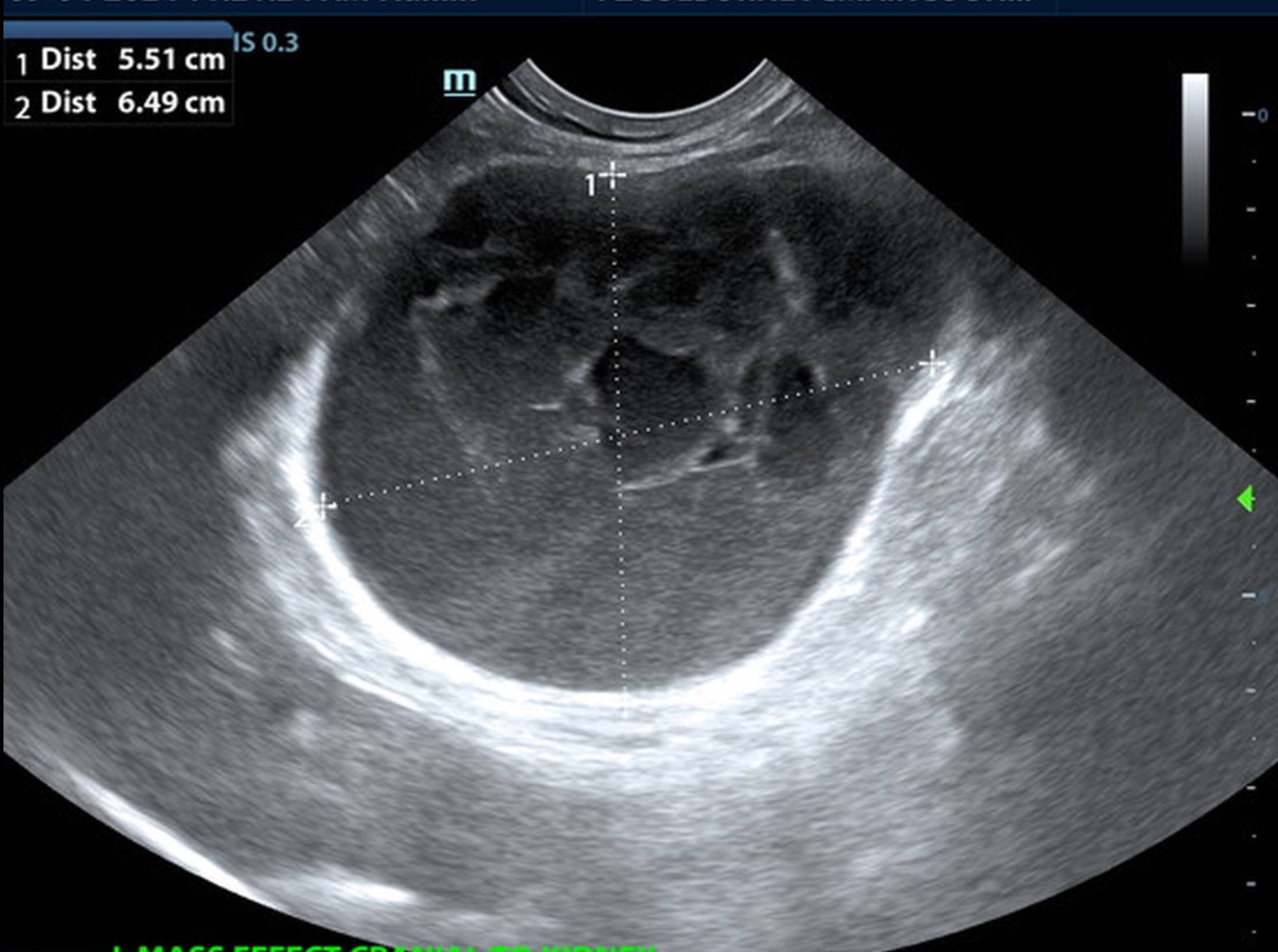
Radiographs: 3-view rads showed circular mid-abdominal mass with soft tissue opacity. Both kidneys visualized independently from the mass. Loss of serosal detail cranial/ventral abdomen (effusion?)


Image Interpretation

A 6.5 cm hypoechoic abscess was noted in the region of the left pancreatic limb with hyperechoic granulation bed and regional inflammation. The abscess was largely non-vascular. One minor portion of color flow uptake noted within the abscess.

Renal: some age-related loss of curvilinear patterns regarding the capsule and C/M junction. The cortices presented largely uniform texture with some increased echogenicity expected for his age patient.

ULTRASONOGRAPHIC FINDINGS

  • Large left-sided pancreatic abscess.
  • Age related renal changes.

Comments

Rapid surgical intervention recommended. Ultrasound guided drainage could be considered. However, there is significant potential for leakage, given the turgid pattern of the abscessation. The remainder of the abdomen was unremarkable other than age related changes. Culture and sensitivity indicated. No evidence nor suspicion of neoplasia.

Patient Information

Gender : Male, Neutered
Species : Feline
Type of Imaging : Ultrasound

Clinical Signs

  • Anorexia
  • Lethargy
  • Vomiting

Exam Finding

  • Abdominal Pain
  • Palpable mass

Images

Clinical Signs

  • Anorexia
  • Lethargy
  • Vomiting
Skip to content