Intrahepatic Shunt, suspect Central Divisional or Right Divisional, in a 8-month-old, Intact Male, DSH Cat

Case Study

Intrahepatic Shunt, suspect Central Divisional or Right Divisional, in a 8-month-old, Intact Male, DSH Cat

Presented for drooling, lethargy, ataxia/falling over. Marked bile acid (pre- and post-prandial) elevation.

PE: Occasional tremors in exam room, O notes ataxia at home. Occasionally falls over on side and can’t get up but seems mentally aware.

Rads: Microhepatica, Loss of serosal detail, Large amount of feces in colon

Labwork: Bile acids Preprandial: 110 H (0-6.9) // BA Post prandial: 122.6 H (0-14.9) CBC: RBC 12.27H, Hct 41.4%, MCV 34 L (39-56),  MCH 11.3 L (12.6-16.5), Lymph 7.4 H (0.85-5.85) Chem: Gluc 131-N, Cr 0.7 L (0.9-2.3), SDMA 3-N, BUN  19-N, Alb 3.4-N, Chol 97-N, ALT 38-N, AST 30 – N, ALP 96 H (12-59), GGT < 1 – N

Sonographic Differential Diagnosis

Suspect intrahepatic shunt, central divisional or right divisional.


Image Interpretation

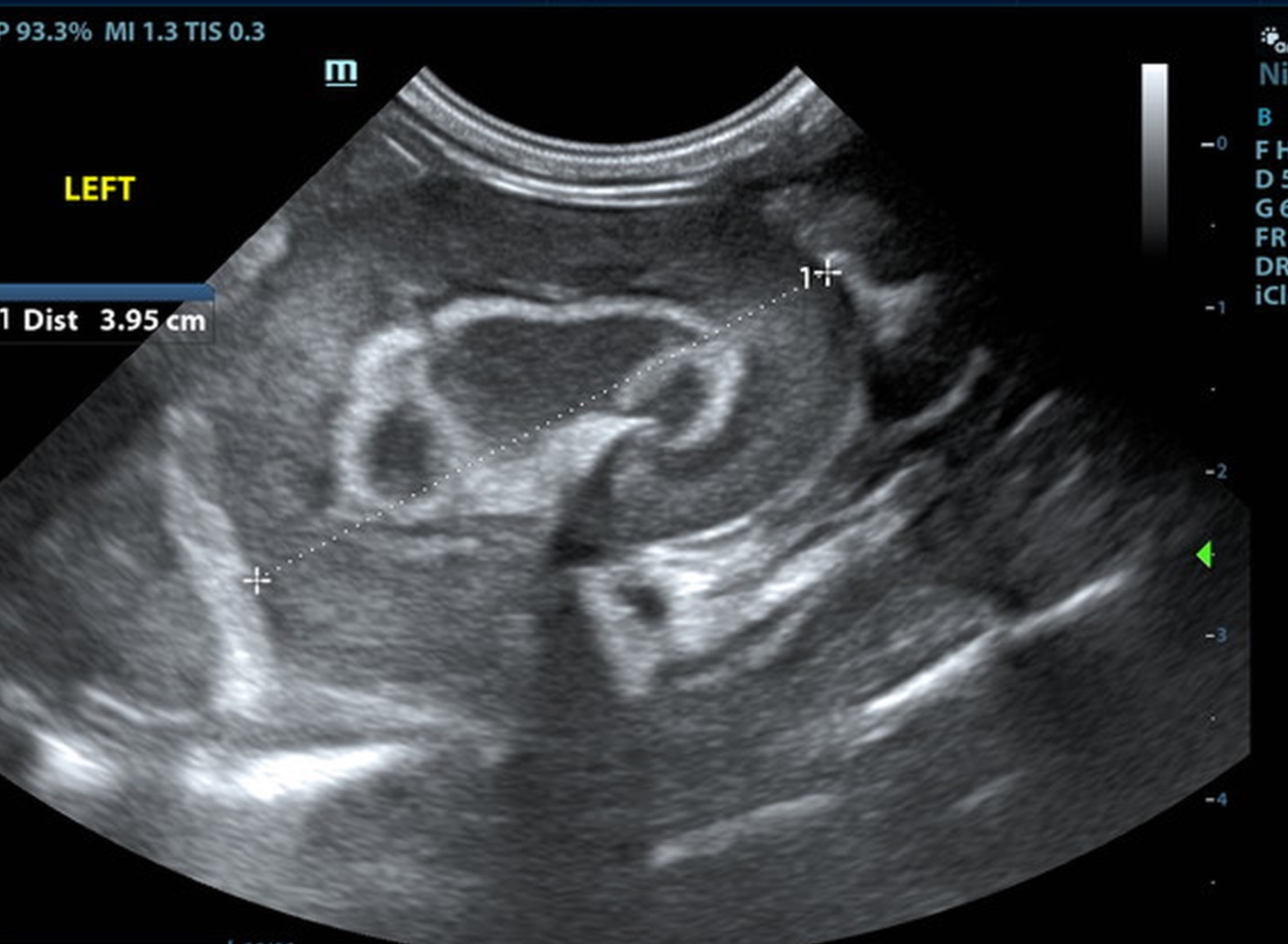
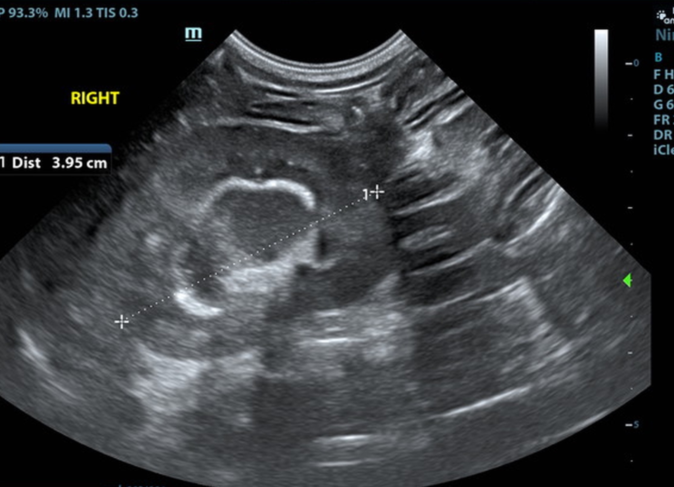
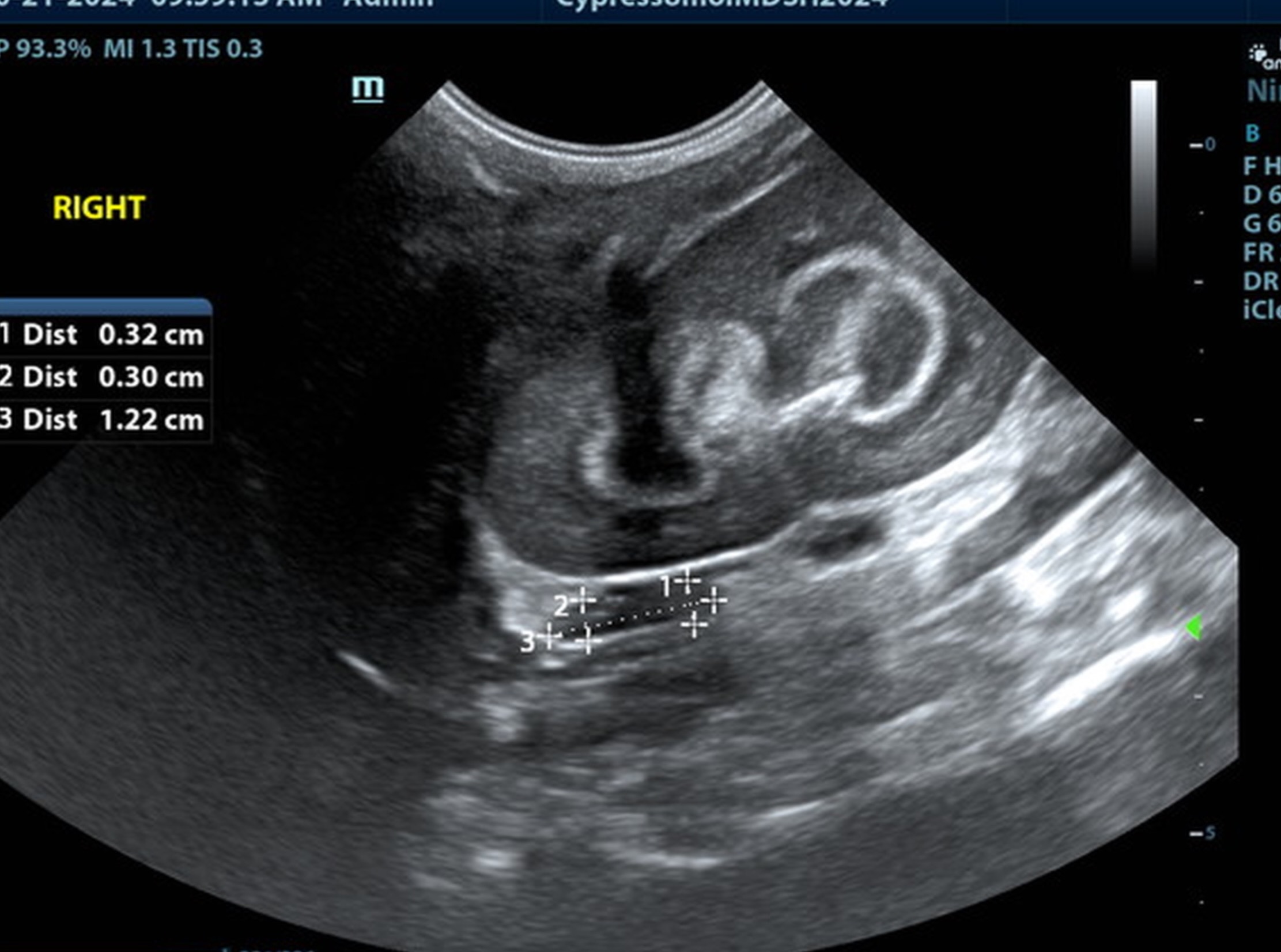
The kidneys were swollen with hyperechoic, mineralized medullary rim sign. The right kidney measured 4.0 cm. The left kidney measured 3.95 cm.

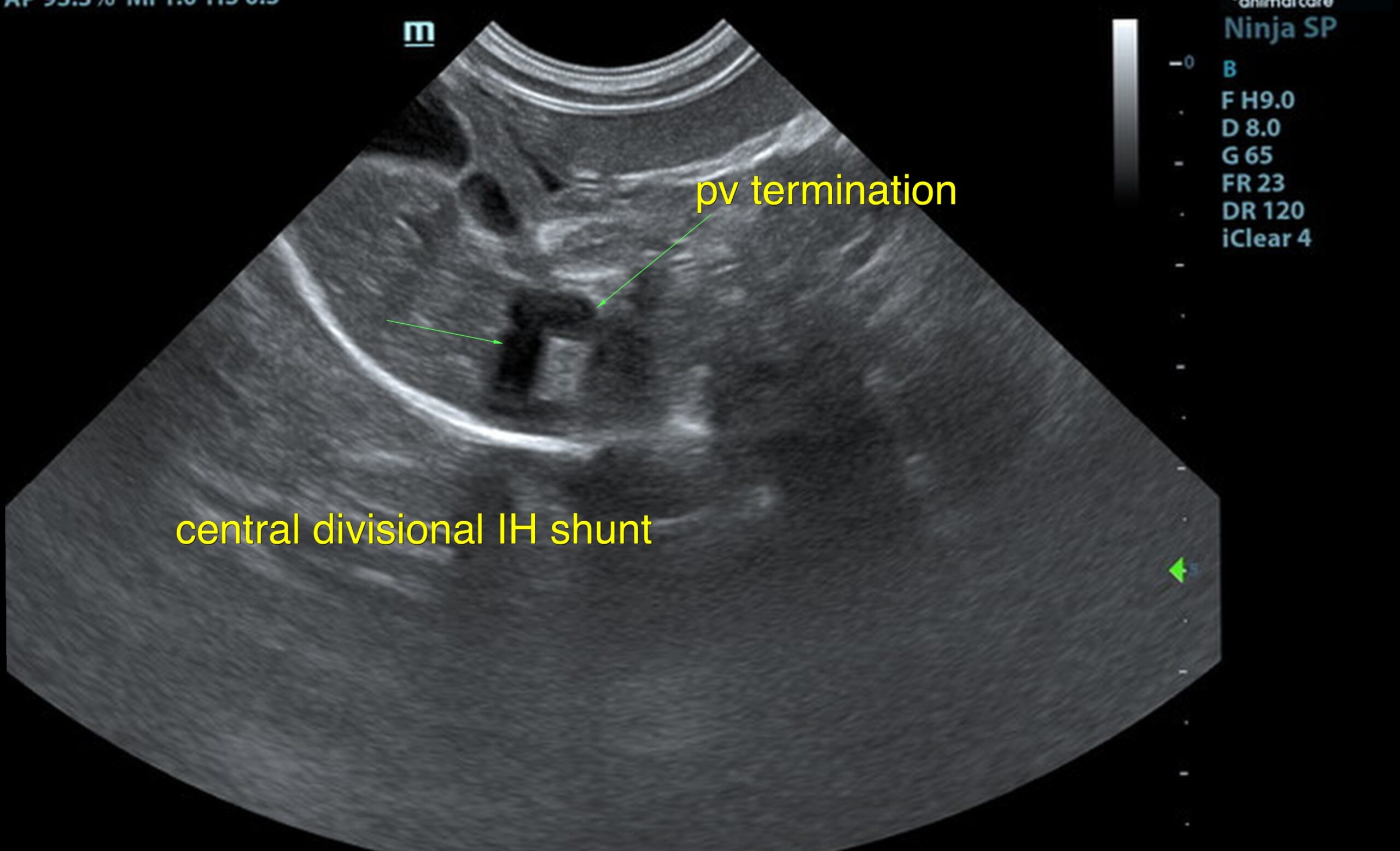
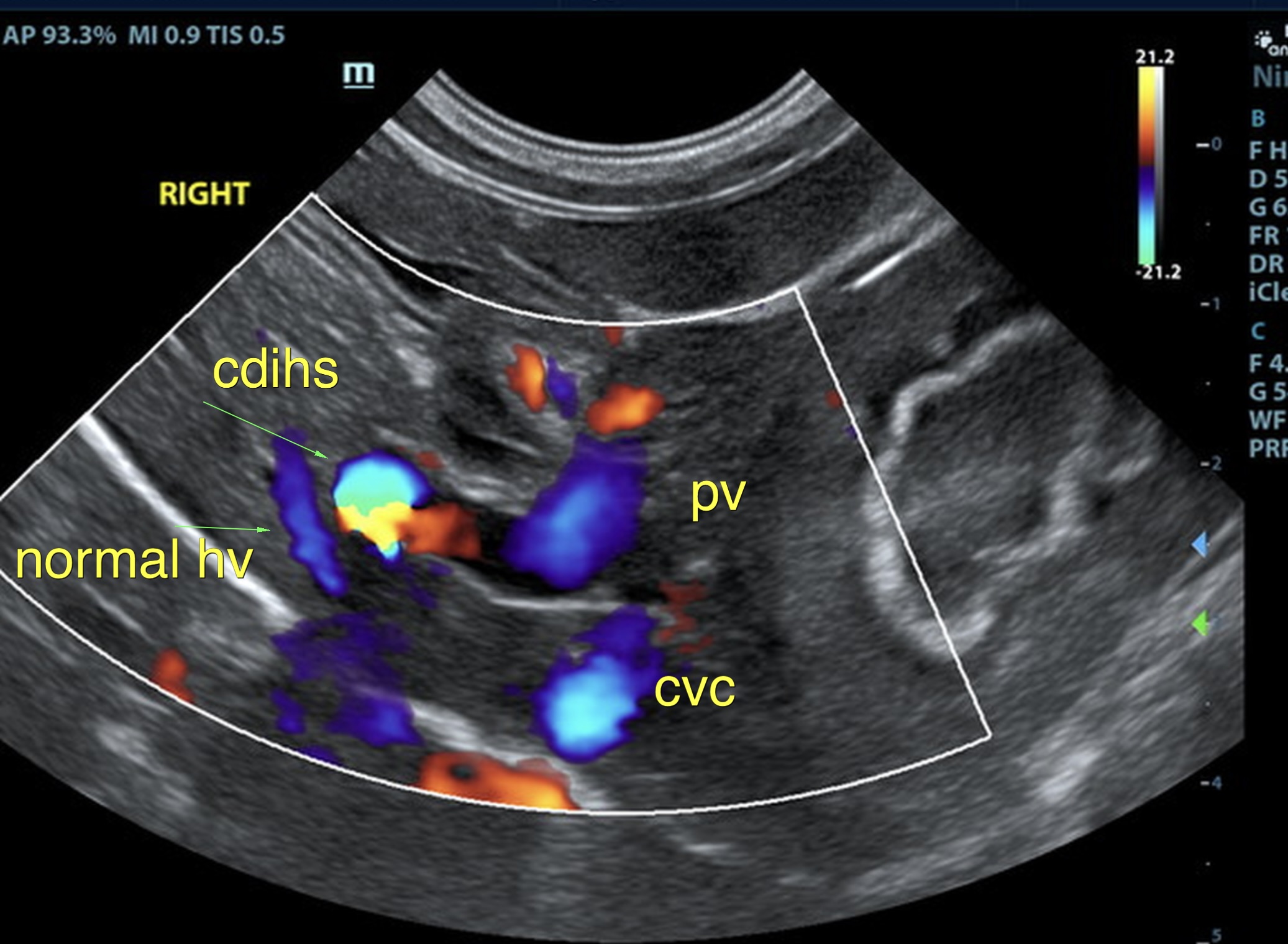
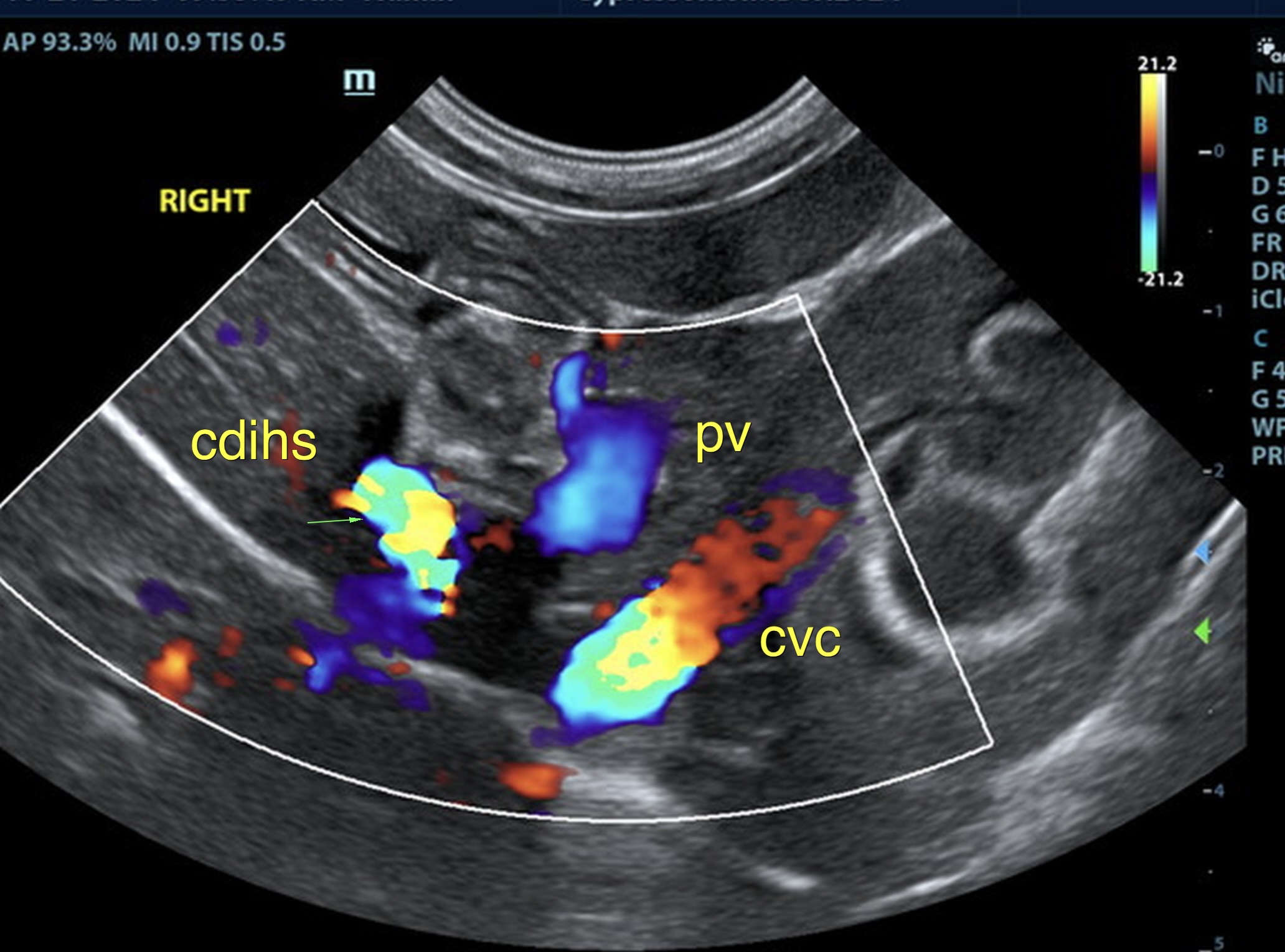
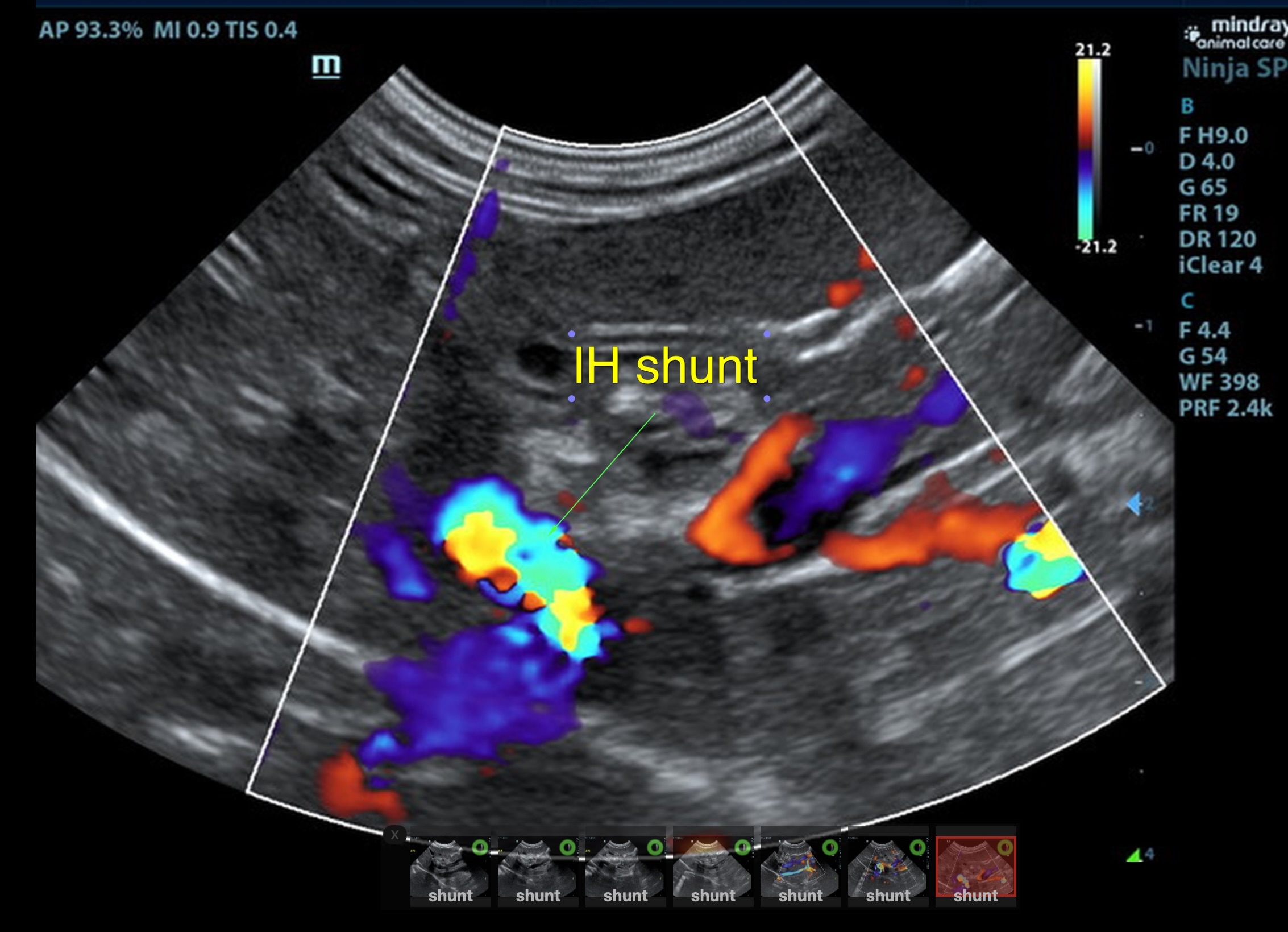
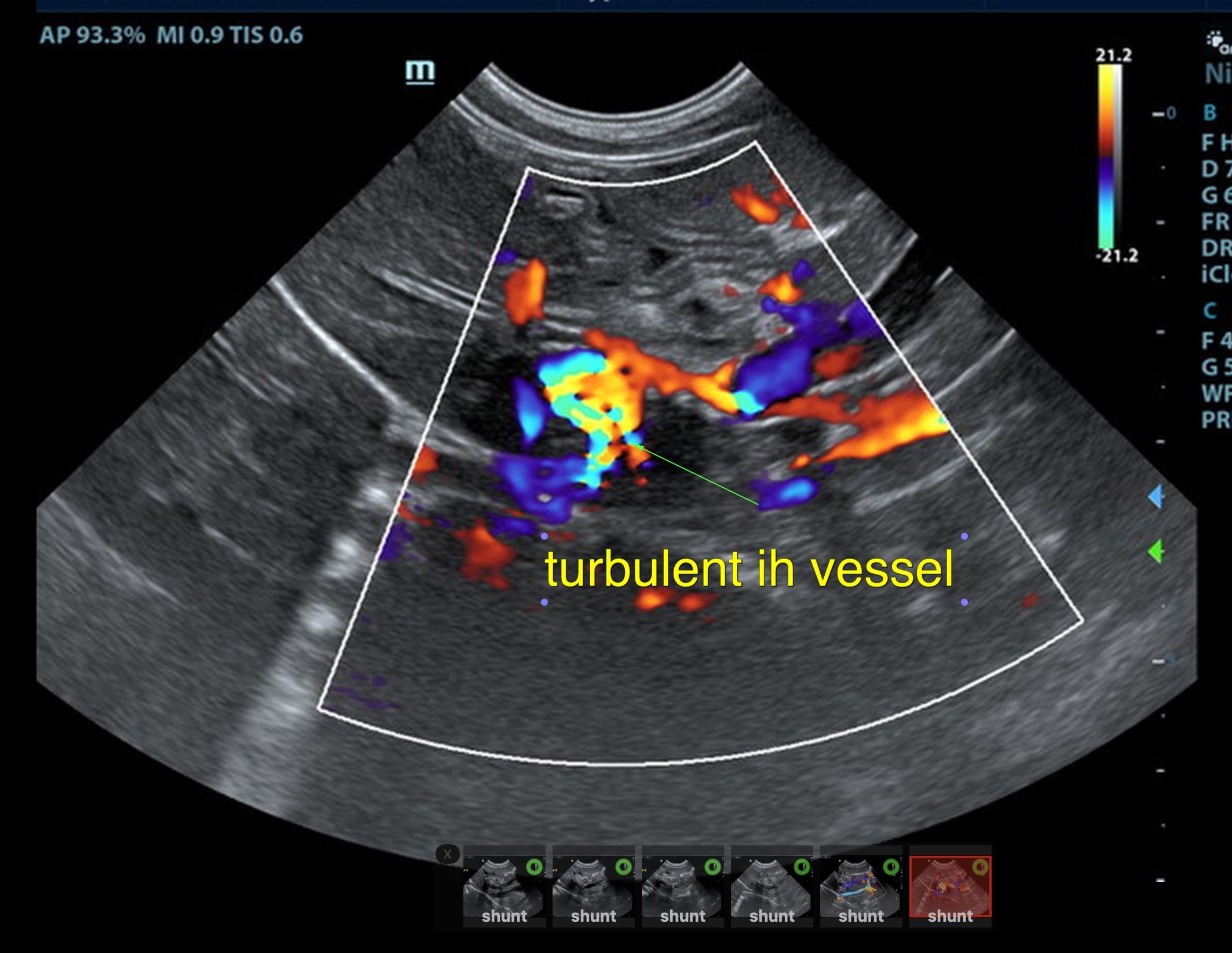
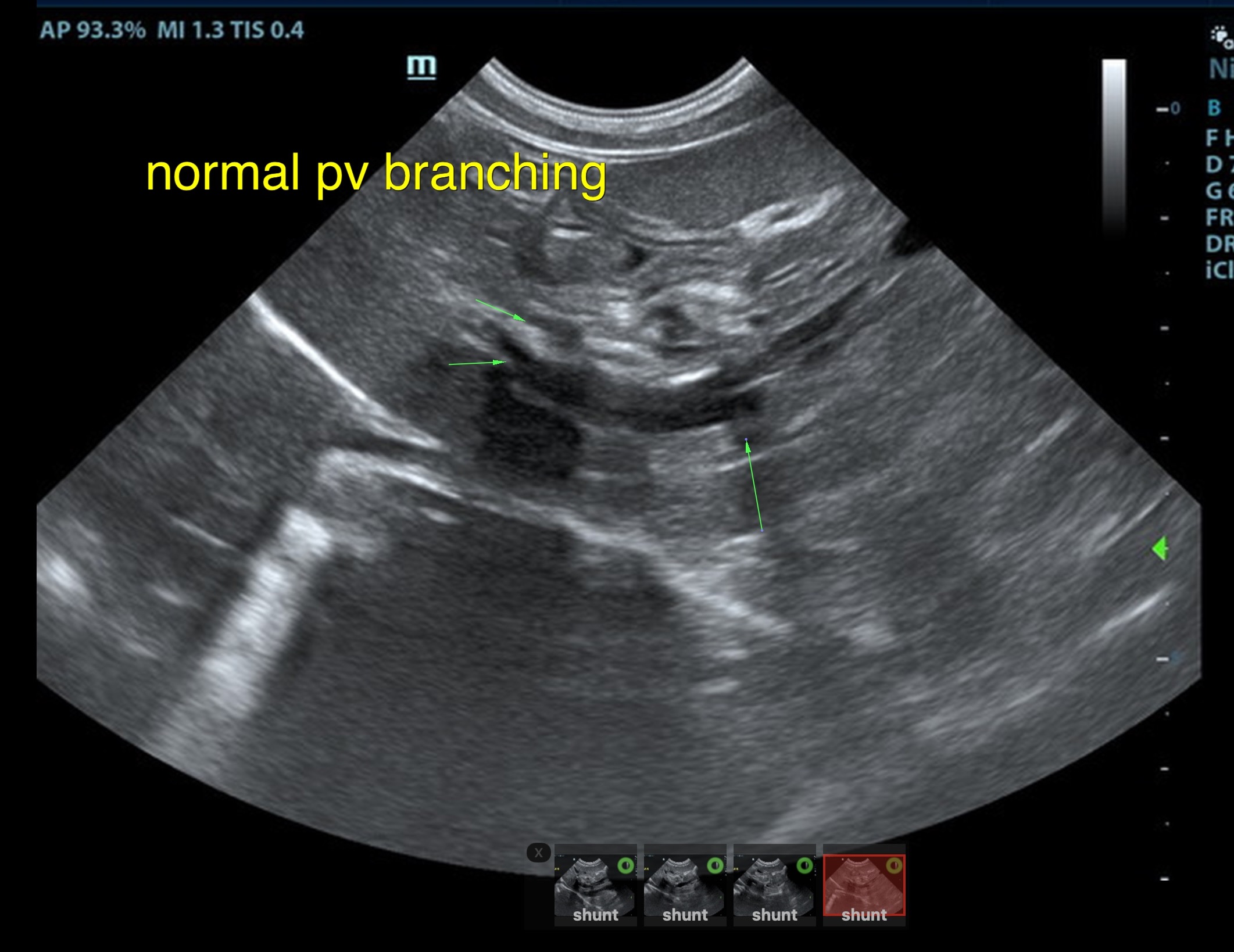
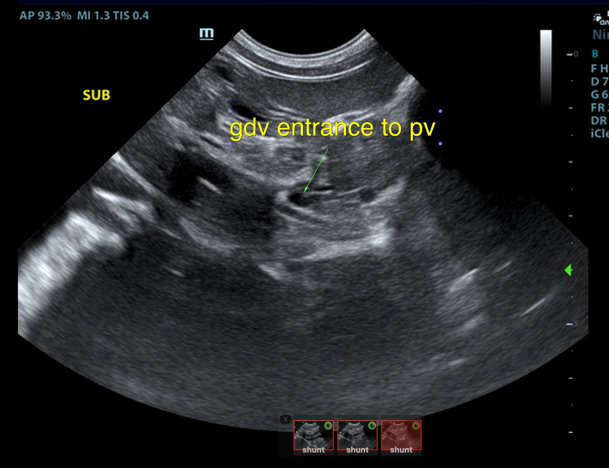
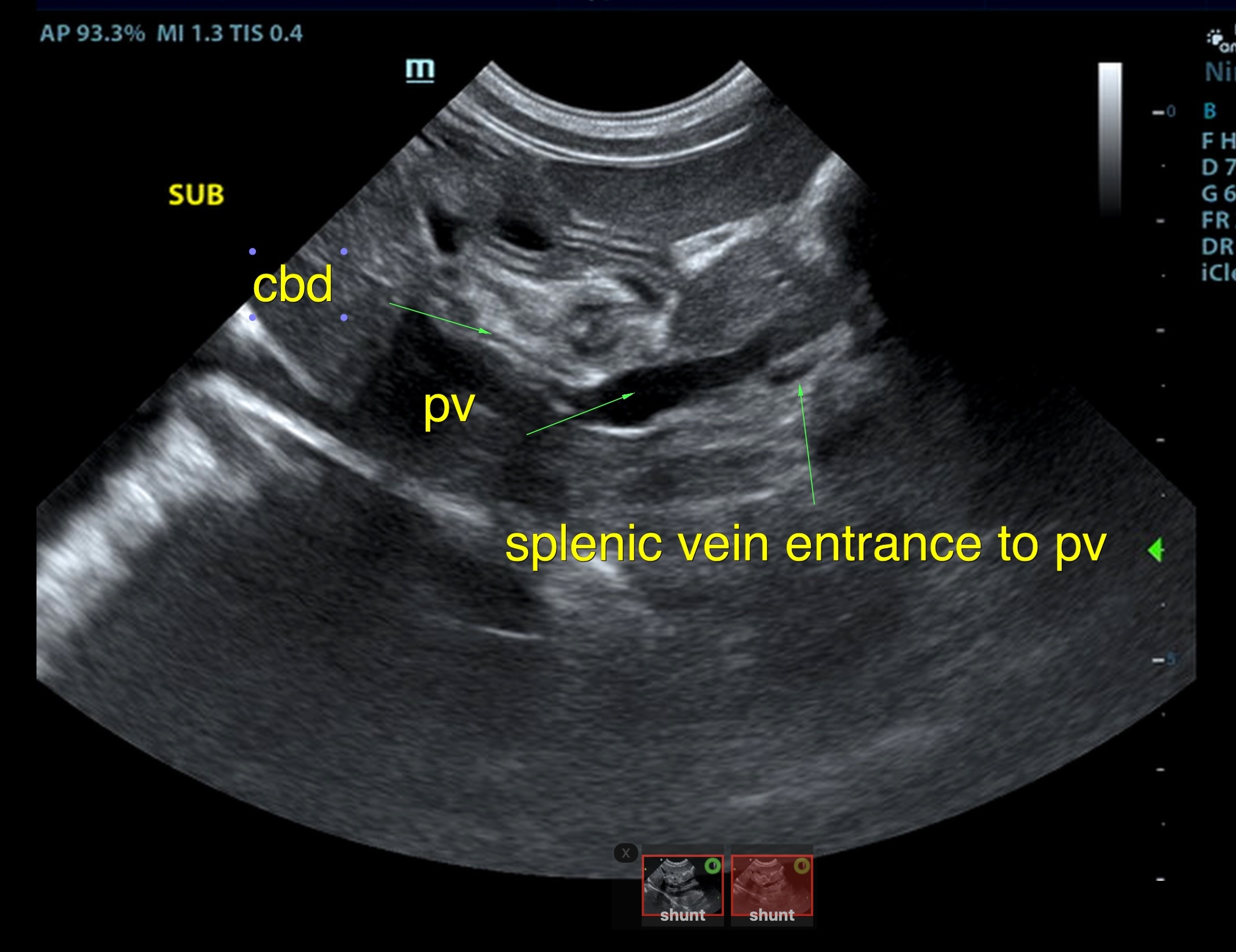
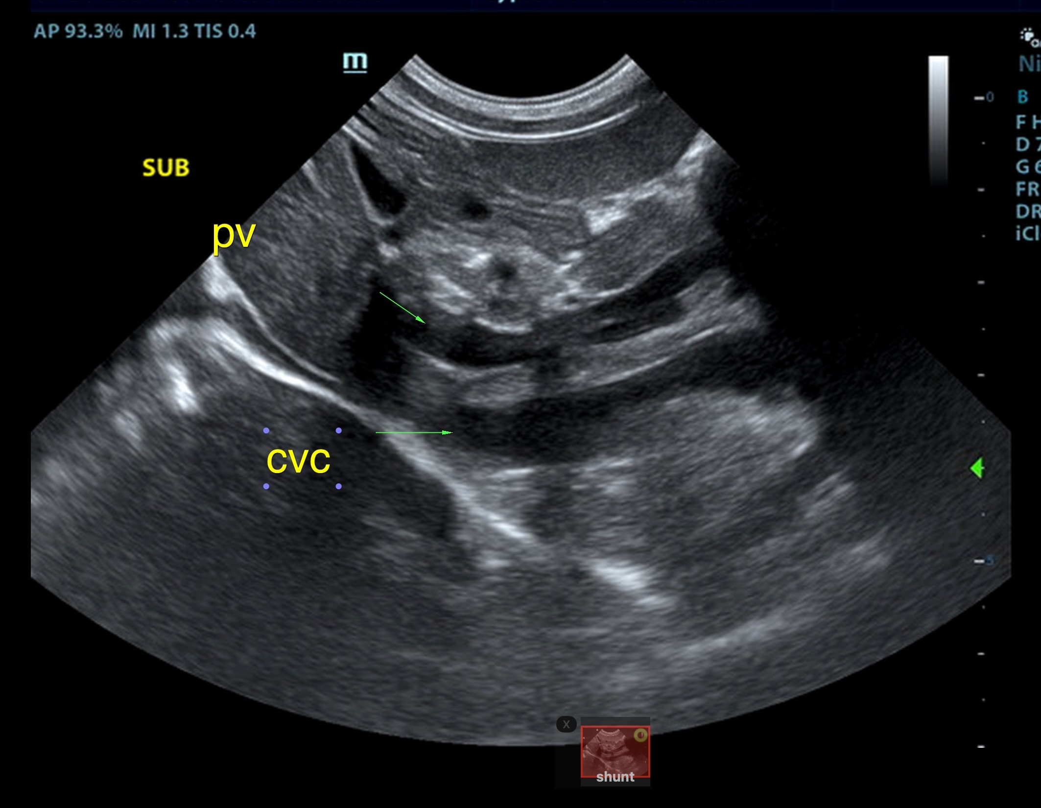
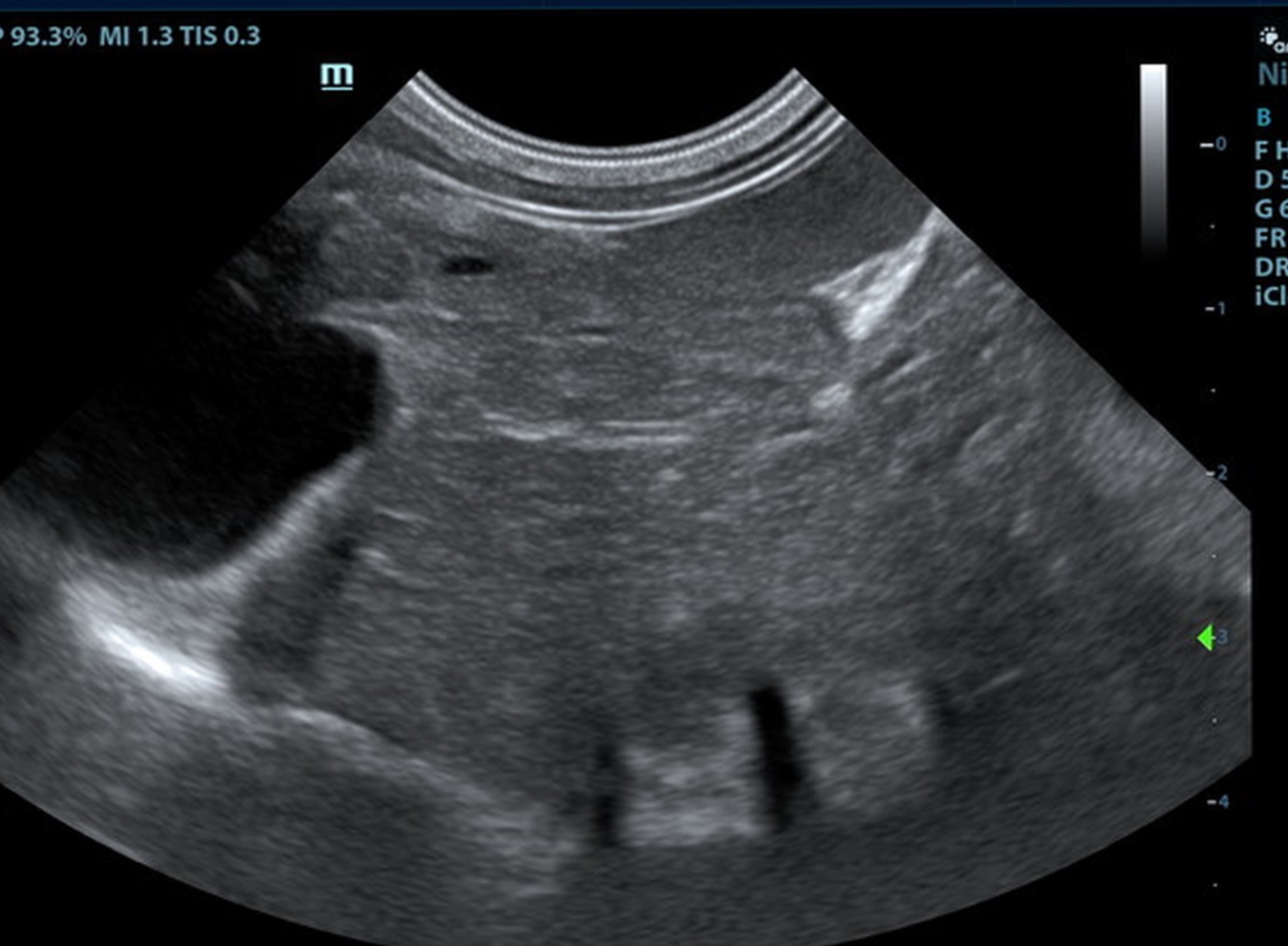
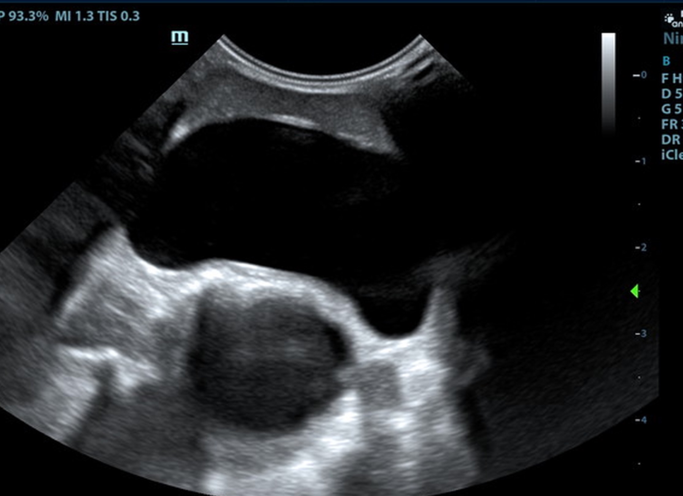
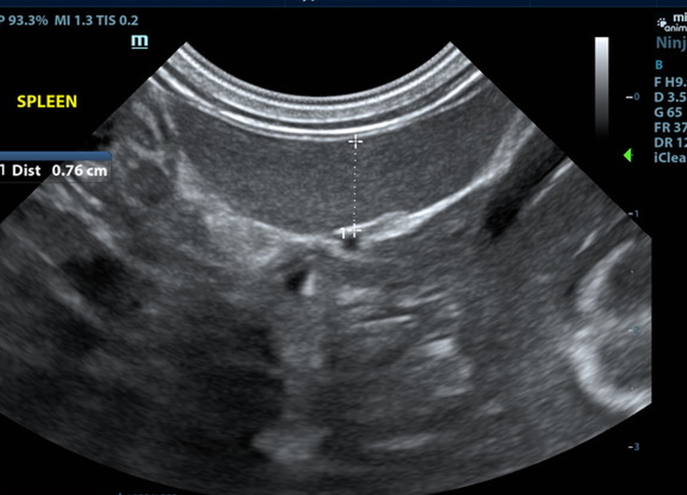
The liver was subnormal in size, yet the portal vein and vena cava ratio was 1:1. The portal veins were subnormal in size and measured 0.34 cm. The vena cava was enlarged and measured 0.34 cm. The vena cava was enlarged and measured 0.72 cm, aorta measured 0.4 cm. The branching of the portal vein appeared to be normal and of adequate volume. The portal vein and vena cava measured 0.5 cm each in the extrahepatic space. The splenic vein entry into the portal vein and gastroduodenal vein entry into the portal vein appear to be normal. There was one turbulent vessel in the region of the central branch of the portal vein, which may represent an intrahepatic shunt, but this could not be confirmed. The width of the shunt is approximately 0.76 cm. This is in position of central divisional shunt; however, right divisional origin cannot be completely ruled out. The gallbladder presented acceptably thin walls with primarily anechoic content. The cystic and common bile ducts were normal.

Suspect intrahepatic shunt, central divisional or right divisional.

Medullary rim kidneys, secondary to abnormal biurate metabolism and renal swelling.

No current bladder calculi.


Comments

CT evaluation with contrast is indicated. Vascular occlusion would be necessary in this patient. Referral for interventional planning and CT confirmation and interventional planning is indicated. Internal medicine consult could be utilized until interventional occlusion can occur.

Clinical Differential Diagnosis

portosystemic shunt

Patient Information

Gender : Male, Intact
Species : Feline
Type of Imaging : Ultrasound

Clinical Signs

  • Ataxia
  • Lethargy

Exam Finding

  • Microhepatica
  • Tremors

Images

Clinical Signs

  • Ataxia
  • Lethargy

Special Testing

  • bile acids pre-/post-prandial
Skip to content